A Science teacher by trade, I've also been known to be found teaching Maths and PE! However, strange as it may seem, my real love is designing resources that can be used by other teachers to maximise the experience of the students. I am constantly thinking of new ways to engage a student with a topic and try to implement that in the design of the lessons.
A Science teacher by trade, I've also been known to be found teaching Maths and PE! However, strange as it may seem, my real love is designing resources that can be used by other teachers to maximise the experience of the students. I am constantly thinking of new ways to engage a student with a topic and try to implement that in the design of the lessons.
This detailed lesson has been planned to cover the content of specification point 14.1 (e) of the CIE International A-level Biology specification which states that students should be able to describe the gross structure of the kidney and the detailed structure of the nephron. The lesson was designed at the same time as the other lessons in this topic on ultrafiltration, selective reabsorption and osmoregulation so that a common theme runs throughout and students can build their knowledge up gradually and develop a deep understanding of this organ.
Students will come to recognise the renal cortex and renal medulla as the two regions of the kidney and learn the parts of the nephron which are found in each of these regions. Time is taken to look at the vascular supply of this organ and specifically to explain how the renal artery divides into the afferent arterioles which carry blood towards the glomerulus and the efferent arterioles which carry the blood away. The main task of the lesson challenges the students to relate structure to function. Having been introduced to the names of each of the parts of the nephron, they have to use the details of the structures found at these parts to match the function. For example, they have to make the connection between the microvilli in the PCT as a sign that this part is involved in selective reabsorption.
This lesson has been designed for students studying on the CIE International A-level Biology course
This is a detailed and engaging lesson which has been designed to cover specification points 14.1 (a, b and c) of the CIE International A-level Biology specification which states that students should be able to explain the importance of homeostasis and the roles of negative feedback and the communication systems in this control.
As homeostasis is a topic met at GCSE, this lesson has been written to build on this knowledge as well as to check on their prior knowledge of earlier A-level topics such as osmosis when considering blood water potential. Discussion points are written into the lesson at regular intervals to encourage the students to consider why a particular process or method takes place and understanding checks allow them to assess their progress. Students will recall how body temperature, blood water potential and blood glucose concentration are maintained within strict limits and the importance of these systems are looked into in detail. They will also learn that carbon dioxide concentration and blood pressure are aspects that are controlled in the body and key terminology such as chemoreceptors and baroreceptors are used throughout so that students are confident with the meaning when met later in the module. The key components of the control system are recalled and then time is taken to focus on the cell signalling that occurs between the coordination centre and the effectors. Students will learn to associate the response with either the use of the neuronal or hormonal system. The final part of the lesson looks at the importance of negative feedback in reversing the change in order to bring it back to the optimum and the differences to positive feedback are also explored.
This lesson has been written for students who are studying the CIE International A-level Biology course and ties in well with the other uploaded lessons on this topic such as those on the kidney
This is a fully-resourced lesson that covers the content of specification point 15.1 (k) of the CIE International A-level Biology specification which states that students should be able to explain the sliding filament model of muscular contraction. The wide range of activities included in the lesson will engage and motivate the students whilst the understanding and previous knowledge checks will not only allow them to assess their progress but also challenge them to make links to other Biology topics.
The start of the lesson is designed to encourage the students to consider how a sarcomere can narrow but the lengths of the myofilaments can remain the same. In doing so, they will be introduced to the idea of the sliding filament model and the main task of the lesson involves the formation of a bullet point description of this model where one event is the trigger for the next. Time is taken during this section to focus on the involvement of the calcium ions but also ATP and the idea of the sources of this molecule, including creatine phosphate, are discussed in more detail later in the lesson. The final part of the lesson involves students having to apply their knowledge by describing the effect on muscle contraction when a part of a structure is unable to function correctly.
This lesson has been designed for students studying the CIE International A-level Biology course and ties in well with the other uploaded lessons on this topic, particularly the lesson which covers the ultrastructure of striated muscle
This concise and engaging lesson has been designed to cover specification point 15.1 (j) of the CIE International A-level Biology specification which states that students should be able to describe the ultrastructure of striated muscle with particular reference to sarcomere structure. The wide range of key terms and regions are introduced in a fun and memorable way using a variety of activities that include quiz competitions and then understanding checks are used throughout to assess their progress and ensure that any misconceptions are addressed. Connections are made to the upcoming topic of the sliding filament model as the students discover that despite the shortening of the sarcomere (and I band and H zone) during contraction, the fact that the A band remains the same length shows how the filaments slide over each other. The two main tasks of the lesson challenge the students to label a diagram of a sarcomere and then the microscope image as shown in the cover picture.
This lesson has been designed to tie in well with the other uploaded lessons that cover the content of topic 15.1 of the CIE International A-level Biology course which is the control and coordination in mammals
This concise, fully-resourced lesson covers the content of specification point 15.1 (i) of the CIE International A-level Biology specification which states that students should be able to describe the roles of the neuromuscular junction, transverse tubules and sarcoplasmic reticulum in the stimulation of the contraction of striated muscle. Due to a number of similarities between these structures and cholinergic synapses, this lesson uses prior knowledge of these connections between neurones to build a good understanding of the junctions. Students will discover that the events that occur at an axon tip mirror those which happen at the pre-synaptic bulb and this is then developed to look at the differences in terms of the events once the acetylcholine has bound to its receptor sites. There is a focus on the structure of the sarcolemma and time is taken to explain how the action potential is passed from this membrane to the transverse tubules in order to stimulate the release of calcium ions from the sarcoplasmic reticulum. As a result, this lesson ties in nicely with the following lesson on the contraction of skeletal muscle and students will be able to link the binding to troponin in that lesson to the release of these ions from this lesson.
Both of the main tasks of the lesson have been differentiated so that students of all abilities can access the work and make progress.
This lesson has been designed for those students studying on the CIE International A-level Biology course and ties in well with the other uploaded lessons on topic 15.1 (Control and coordination in mammals)
This fully-resourced lesson covers the content of specification points 15.1 (g and h) of the CIE international A-level Biology specification that states that students should be able to describe the structure of a cholinergic synapse and outline their roles in the nervous system. The majority of the lesson uses the cholinergic synapse as the example but other neurotransmitters are considered at the end of the lesson to provide the students with a wider view of this topic.
The lesson begins by using a version of the WALL (as shown in the cover image) which asks the students to group 12 words into three groups of 4. Not only will this challenge their prior knowledge from topics earlier in this module but it will also lead to the discovery of four of the structures that are found in a synapse. Moving forwards, students are introduced to acetylcholine as the neurotransmitter involved at cholinergic synapses and they will start to add labels to the structures found in the pre-synaptic bulb. Time is taken to focus on certain structures such as the voltage gated channels as these types of channel were met previously when looking at the depolarisation of a neurone. There is plenty of challenge and discovery as students are pushed to explain why organelles like mitochondria would be found in large numbers in the bulb. With this process being a cascade of events, a bullet point format is used to ensure that the key content is taken in by the students and again key points like exocytosis and the action of acetylcholinesterase are discussed further. The final part of the lesson challenges the application aspect of the subject as students are introduced to unfamiliar situations in terms of synapses with new drugs like MDMA and are asked to work out and explain how these affect the nervous transmission.
Understanding checks and prior knowledge checks are included throughout the lesson so that students can not only assess their progress against the current topic but also see whether they can make links to earlier topics.
This lesson has been designed for students studying the CIE International A-level Biology course and ties in with the other uploaded lessons on the topics of 15.1 (Control and coordination in mammals)
This lesson has been written to cover the detail of specification point 15.1 (f) of the CIE International A-level Biology specification which states that students should be able to explain the importance of myelination. A wide range of activities have been written into this resource to maintain the motivation of the students whilst ensuring that the detail is covered in depth. Interspersed with the activities are understanding checks and prior knowledge checks to allow the students to not only assess their understanding of the current topic but also challenge themselves to make links to earlier topics such as the movement of ions across membranes and biological molecules. Time at the end of the lesson is also given to future knowledge such as the involvement of autonomic motor neurones in the stimulation of involuntary muscles.
Over the course of the lesson, students consider the structure of the myelin sheath and specifically how the insulation is not complete all the way along which leaves gaps known as the nodes of Ranvier which allow the entry and exit of ions. Saltatory conduction is poorly explained by a lot of students so time is taken to look at the way that the action potential jumps between the nodes and this is explained further by reference to local currents. The rest of the lesson focuses on the other two factors which are axon diameter and temperature and students are challenged to discover these two by focusing on the vampire squid.
This lesson has been designed for students studying the CIE International A-level Biology course and the other part of this specification point which covers the refractory period was explained in the previous lesson on the transmission of the action potential
This is a highly detailed and engaging lesson that covers the detail of specification point 15.1 (e) of the CIE International A-level Biology specification which states that students should be able to describe and explain the transmission of an action potential in a myelinated neurone. This topic is commonly assessed in the terminal exams so a lot of time has been taken to design this resource to include a wide range of activities that motivate the students whilst ensuring that the content is covered in the depth of detail that will allow them to have a real understanding. Interspersed within the activities are understanding checks and prior knowledge checks to enable the students to not only assess their progress against the current topic but also to challenge themselves on the links to earlier topics such as methods of movements across cell membranes. There are also a number of quiz competitions which are used to introduce key terms and values in a fun and memorable way and discussion points to encourage the students to consider why a particular process or mechanism occurs.
Over the course of the lesson, the students will learn and discover how the movement of ions across the membrane causes the membrane potential to change. They will see how the resting potential is maintained through the use of the sodium/potassium pump and potassium ion leakage. There is a real focus on depolarisation to allow students to understand how generator potentials can combine and if the resulting depolarisation then exceeds the threshold potential, a full depolarisation will occur. At this point in the lesson students will discover how the all or nothing response explains that action potentials have the same magnitude and that instead a stronger stimulus is linked to an increase in the frequency of the transmission. The rest of the lesson challenges the students to apply their knowledge to explain how repolarisation and hyperpolarisation result and to suggest advantages of the refractory period for nerve cells.
This lesson has been designed for students studying the CIE International A-level Biology course and ties in nicely with other uploaded lessons which cover the content of topic 15.1 (Control and coordination in mammals)
This is a fully-resourced lesson which covers the detail of specification point 15.1 (b) of the CIE International A-level Biology specification which states that students should be able to describe the structure of a sensory and a motor neurone. The PowerPoint has been designed to contain a wide range of activities that are interspersed between understanding and prior knowledge checks that allow the students to assess their progress on the current topics as well as challenge their ability to make links to topics from earlier in the modules. Quiz competitions like SAY WHAT YOU SEE are used to introduce key terms in a fun and memorable way.
The students will be able to compare these neurones based on their function but also distinguish between them based on their structural features. Time is taken to look at the importance of the myelin sheath that is present in both neurones. Students will be introduced to the need for the entry of ions to cause depolarisation and will learn that this is only possible at the nodes of Ranvier when there is a myelin sheath. Key terminology such as saltatory conduction is introduced and explained and the lesson concludes with the introduction of the different types of motor neurones based on the type of muscle which they innervate.
This lesson has been designed for students studying on the CIE International A-level Biology course and ties in well with the other uploaded lessons which cover the content of topic 15.1 (Control and coordination in mammals) .
This bundle contains 17 fully-resourced and detailed lessons that have been designed to cover the content of topic 6 of the AQA A-level Biology specification which concerns the responses of organisms to stimuli. The wide range of activities included in each lesson will engage the students whilst the detailed content is covered and the understanding and prior knowledge checks allow them to assess their progress on the current topic as well as challenging them to make links to other related topics. Most of the tasks are differentiated to allow differing abilities to access the work and be challenged.
The following sub-topics are covered in this bundle of lessons:
The role of sensory receptors as outlined by the Pacinian corpuscle
The human retina as a sensory receptor
The differences in rods and cones that enable different sensitivity to light, colour and visual acuity
The roles of the SAN, AVN, Bundle of His and Purkyne fibres in the conduction system of the heart
The control of heart rate
The structure of a myelinated motor neurone
The factors that affect the speed of conduction of an impulse
The generation and transmission of nerve impulses
The transmission at a cholinergic synapse and a neuromuscular junction
Summation
The contraction of skeletal muscles
The structure and properties of slow and fast skeletal muscle fibres
The principles of homeostasis including negative feedback systems
The control of blood glucose concentration by the controlled release of insulin and glucagon
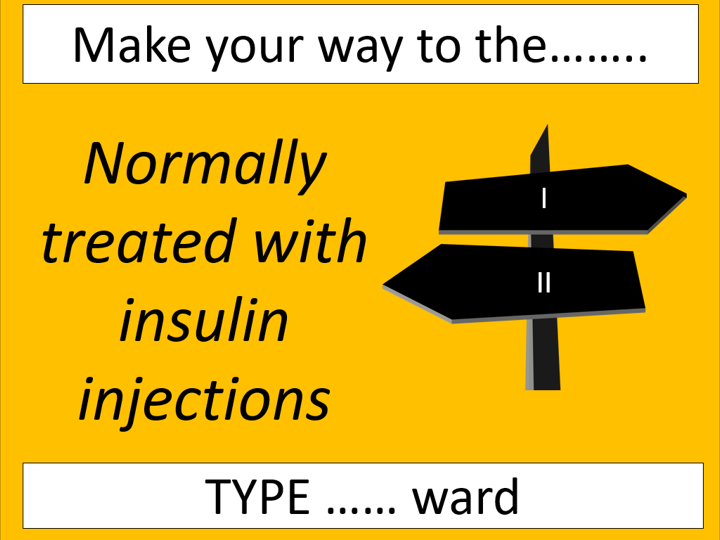
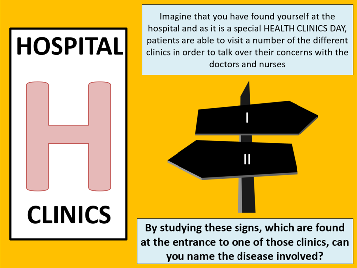
The causes and control of diabetes type I and II
The gross structure of the kidney
The detailed structure of the nephron
The production of glomerular filtrate
The reabsorption of glucose and water in the PCT
The role of the hypothalamus, posterior pituitary and ADH in osmoregulation
This is one of the 8 topics which have to be covered over the length of the 2 year course and therefore it is expected that the teaching time for this bundle will be in excess of 2 months
If you want to see the quality of the lessons before purchasing then the lessons on saltatory conduction, the contraction of skeletal muscles and ultrafiltration are free resources to download
This engaging and fully-resourced lesson covers the content of specification points 5.1.4 (e and f) of the OCR A-level Biology A specification which states that students should be able to demonstrate and apply an understanding of the differences between diabetes mellitus type I and II and the potential treatments of this disease. The lesson has been designed to take place in a diabetes clinic where students will be challenged to perform a number of roles such as diagnosing a patient with either type I or II and to write a letter to this patient explaining how the disease was caused and any treatments that will be recommended to control the disease. It has been planned to build on the knowledge that they will have of these diseases from GCSE and links are made to other A-level topics such as the beta cells of the pancreas which they considered during the lesson on the control of blood glucose concentration. The final part of the lesson looks at the potential treatments which include the genetic modification of bacteria. This topic is covered in greater detail in module 6.1.3 so this section of the lesson focuses on the enzymes involved as well as the plasmid DNA from a bacterial cell.
This lesson has been designed for students studying the OCR A-level Biology A course and runs alongside the uploaded lesson on the control of blood glucose concentration as well as the other lessons that have been added for module 5.1.4
This highly detailed, fully-resourced lesson has been designed to cover the content of specification point 5.1.4 (d) of the OCR A-level Biology A specification which states that students should be able to demonstrate and apply an understanding of the regulation of blood glucose concentration. There is focus on the negative feedback mechanisms that release insulin or glucagon and the role of the liver. It challenges the students recall of the control of insulin release from the beta cells which was taught in an earlier lesson.
A wide range of activities will maintain motivation and engagement whilst the content is covered in detail to enable the students to explain how the receptors in the pancreas detect the concentration change and how the hormones attaching to receptor sites on the liver triggers a series of events in this effector organ. This is a topic which has a huge amount of difficult terminology so time is taken to look at all of the key words, especially those which begin with the letter G so students are able to use them accurately in the correct context. The action of adrenaline is also considered and linked to the breakdown of glycogen to glucose during glycogenolysis.
This lesson has been written for students studying on the OCR A-level Biology A course and ties in with the lesson on the differences between type I and II diabetes mellitus as well as the human endocrine system
This is a highly-detailed and fully-resourced lesson which covers the detail of specification point 5.1.2 (d) of the OCR A-level Biology A specification which states that students should be able to demonstrate and apply an understanding of the roles of the hypothalamus, posterior pituitary, ADH and the collecting duct in the control of the water potential of the blood. Students learnt about the principles of homeostasis and negative feedback in an earlier module, so this lesson acts to build on that knowledge and challenges them to apply their knowledge. A wide range of activities have been included in the lesson to maintain motivation and engagement whilst the understanding and prior knowledge checks will allow the students to assess their progress as well as challenge themselves to make links to other Biology topics.
The lesson begins with a discussion about how the percentage of water in urine can and will change depending on the blood water potential. Students will quickly be introduced to osmoregulation and they will learn that the osmoreceptors and the osmoregulatory centre are found in the hypothalamus. A considerable amount of time is taken to study the cell signalling between the hypothalamus and the posterior pituitary gland by looking at the specialised neurones (neurosecretory cells). Links are made to the topics of neurones, nerve impulses and synapses and the students are challenged to recall the cell body, axon and vesicles. The main section of the lesson forms a detailed description of the body’s detection and response to a low blood water potential. The students are guided through this section as they are given 2 or 3 options for each stage and they have to use their knowledge to select the correct statement. The final task asks the students to write a detailed description for the opposite stimulus and this task is differentiated so those who need extra assistance can still access the work.
This lesson has been written for students studying on the OCR A-level Biology A course and ties in nicely with the other uploaded lessons in module 5.1.2 which include the structure of the nephron, ultrafiltration and selective reabsorption.
This lesson has been written to cover the part of specification point 6.4.3 of the AQA A-level Biology specification which states that students should be able to describe how water and glucose are reabsorbed in the proximal convoluted tubule. It has specifically been designed to build on the knowledge gained in the previous lessons on the structure of the nephron and ultrafiltration.
The lesson begins by challenging the students to recall the substances that are found in the glomerular filtrate so that each of them can be considered over the course of the rest of the lesson. Moving forwards, the first of the numerous discussion points which are included in the lesson is used to get students to predict the component of the filtrate which won’t be found in the urine when they are presented with pie charts from each of these situations. Upon learning that glucose is 100% reabsorbed, along with most of the ions and some of the water, the rest of the lesson focuses on describing the relationship between the structure of the PCT and the function of selective reabsorption. Again, this section begins by encouraging the students to discuss and to predict which structures they would expect to find in a section of the kidney if the function is to reabsorb. They are given the chance to see the structure (as shown in the cover image) before each feature is broken down to explain its importance. Time is taken to look at the role of the cotransporter proteins to explain how this allows glucose, along with sodium ions, to be reabsorbed from the lumen of the PCT into the epithelial cells. The final part of the lesson focuses on urea and how the concentration of this substance increases along the tubule as a result of the reabsorption of some of the water.
This lesson has been designed for students studying on the AQA-A level Biology course and ties in nicely with the other lessons from 6.4.3 as well as the other uploaded lessons from topic 6
This detailed lesson has been written to cover the part of specification point 6.4.3 of the AQA A-level Biology specification which states that students should be able to describe how the structure of the nephron allows for the formation of glomerular filtrate. The aim of the design was to give the students the opportunity to discover the function of ultrafiltration and to be able to explain how the mechanisms found in the glomerulus and the Bowman’s capsule control the movement of small molecules out of the blood plasma. Key terminology is used throughout and students will learn how the combination of the capillary endothelium and the podocytes creates filtration slits that allow glucose, water, urea and ions through into the Bowman’s capsule but ensure that blood cells and plasma proteins remain in the bloodstream. A number of quiz competitions are used to introduce key terms and values in a fun and memorable way whilst understanding and prior knowledge checks allow the students to assess their understanding of the current topic and to challenge themselves to make links to earlier topics. The final task of the lesson challenges the students to apply their knowledge by recognising substances found in a urine sample that shouldn’t be present and to explain why this would cause a problem
This lesson has been written for students studying on the AQA A-level course and ties in nicely with the other kidney lessons on the structure of the nephron, selective reabsorption and osmoregulation
This detailed lesson has been planned to cover the 1st part of specification point 6.4.3 of the AQA A-level Biology specification which states that students should be able to describe the detailed structure of the nephron and understand its role in ultrafiltration, selective reabsorption and osmoregulation. The lesson was designed at the same time as the other lessons in this topic on ultrafiltration, selective reabsorption and osmoregulation so that a common theme runs throughout and students can build up their knowledge gradually in order to develop a deep understanding of this organ.
Students will come to recognise the renal cortex and renal medulla as the two regions of the kidney and learn the parts of the nephron which are found in each of these regions. Time is taken to look at the vascular supply of this organ and specifically to explain how the renal artery divides into the afferent arterioles which carry blood towards the glomerulus and the efferent arterioles which carry the blood away. The main task of the lesson challenges the students to relate structure to function. Having been introduced to the names of each of the parts of the nephron, they have to use the details of the structures found at these parts to match the function. For example, they have to make the connection between the microvilli in the PCT as a sign that this part is involved in selective reabsorption.
This lesson has been designed for students studying on the AQA A-level Biology course
This engaging lesson covers the final details of specification point 6.4.2 of the AQA A-level Biology specification which states that students should be able to describe the causes and control of diabetes mellitus type I and II. The lesson has been designed to take place in a diabetes clinic where students will be challenged to perform a number of roles such as diagnosing a patient with either type I or II and to write a letter to this patient explaining how the disease was caused and any treatments that will be recommended to control the disease. It has been planned to build on the knowledge that they will have of these diseases from GCSE and links are made to other A-level topics such as the beta cells of the pancreas which they considered during the lesson on the control of blood glucose concentration.
This lesson has been designed for students taking the AQA A-level Biology course and runs alongside the uploaded lesson on the control of blood glucose concentration as well as the other lessons that have been added on topic 6
This fully-resourced lesson is highly detailed and in combination with the uploaded lesson on the causes of diabetes type I and II, it covers all of specification point 6.4.2 of the AQA A-level Biology specification which states that students should be able to describe the homeostatic control of blood glucose concentration using negative feedback mechanisms that release insulin or glucagon. A wide range of activities will maintain motivation and engagement whilst the content is covered in detail to enable the students to explain how the receptors in the pancreas detect the concentration change and how the hormones attaching to receptor sites on the liver triggers a series of events in this effector organ. This is a topic which has a huge amount of difficult terminology so time is taken to look at all of the key words, especially those which begin with the letter G so students are able to use them accurately in the correct context. The action of adrenaline is also considered and linked to the breakdown of glycogen to glucose during glycogenolysis.
This lesson has been written for students studying on the AQA A-level Biology course and ties in with the already mentioned lesson on diabetes but also with the other uploaded lessons on topic 6 such as nerve impulses and kidney function